

Prostate cancer is a prevalent form of cancer among individuals assigned male at birth, with significant implications for morbidity and mortality. In recent years, the introduction of a prostate-specific membrane antigen positron emission tomography (PSMA PET) scan has revolutionized the detection and management of prostate cancer. This article explores the key aspects of the PSMA PET test, including its purpose, accuracy, and duration.
Understanding the PSMA PET Test
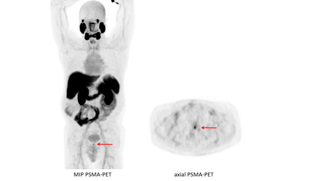
The PSMA PET scan employs a radioactive tracer that targets the prostate-specific membrane antigen (PSMA) protein, which is abundantly present on prostate cancer cells. By injecting the radioactive tracer into the arm, medical professionals can visualize the entire body using a PET scan machine. The PSMA cells light up under the scan, allowing doctors to identify the presence and location of prostate cancer cells, as well as detect metastasis.
Clinical Applications of the PSMA PET Test
Currently, the primary use of the PSMA PET test is to assess the extent of prostate cancer metastasis. It is particularly beneficial for individuals at high or moderate risk of prostate cancer, those with tumors extending beyond the prostate, and those with recurrent cancer post-treatment. The PSMA PET scan aids in determining the spread of cancer, guiding treatment decisions, and monitoring disease progression.
Accuracy of the PSMA PET Test
Studies indicate that the PSMA PET test is approximately 27% more accurate than traditional imaging methods in detecting prostate cancer cells. It demonstrates higher sensitivity and specificity, leading to enhanced detection rates compared to other imaging techniques. The increased accuracy of the PSMA PET scan allows for more precise staging and better understanding of the disease’s extent.
Duration of the PSMA PET Scan
The PSMA PET scan involves a straightforward process. After the injection of the radioactive tracer, individuals typically wait for approximately one hour for the substance to be absorbed by the body. Subsequently, the person lies inside the PET imaging machine for around 20 minutes while the scan takes place. The entire procedure usually lasts a couple of hours, making it a relatively quick and non-invasive imaging test.
The PSMA PET scan has emerged as a game-changing imaging test for the detection and management of prostate cancer. By utilizing the PSMA protein as a target, this innovative technology allows for accurate identification and localization of prostate cancer cells, facilitating improved treatment planning and monitoring. With its higher accuracy compared to traditional imaging methods, the PSMA PET test is transforming the field of prostate cancer diagnosis and staging, offering new hope for patients and healthcare professionals alike.